
Brown Adipose Tissue (BAT) is Brown Fat
Background

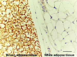
Brown adipose tissue (brown fat) is distinct from white adipose tissue (or white fat). Brown adipose tissue is found in almost all mammals.
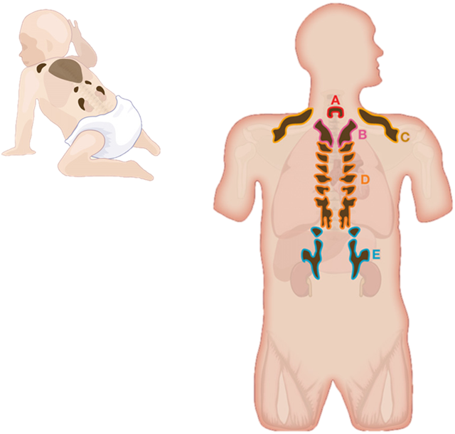
Brown fat is found in highly vascularized deposits in somewhat consistent anatomical locations, such as between the shoulder blades, surrounding the kidneys, the neck and supraclavicular area, and along the spinal cord.
Brown adipose tissue is especially abundant in newborns and in hibernating mammals. It is also present and metabolically active in adult humans, but its prevalence decreases as humans age. Its primary function is thought to be thermoregulation. In addition to heat produced by shivering muscle, brown adipose tissue produces heat by non-shivering thermogenesis because of a unique protein called uncoupling protein 1 or UCP1, which was named thermogenin when originally discovered.
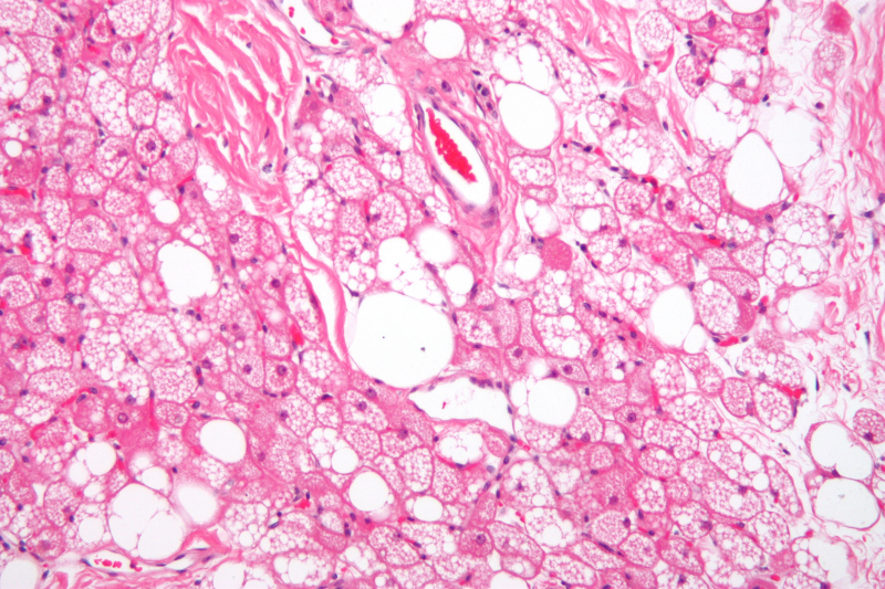
In contrast to white adipocytes (white fat cells), which contain a single lipid droplet, brown adipocytes contain numerous smaller droplets and a much higher number of iron-containing mitochondria, which gives the tissue its color. Brown fat also contains more capillaries than white fat. These supply the tissue with oxygen and nutrients, and distribute the produced heat throughout the body.

The classic population of brown fat cells and muscle cells both seem to be derived from the same population of stem cells in the paraxial mesoderm.
Function
In endotherms, a specialized protein in the mitochondria known as UCP1 (uncoupling protein 1) causes energy to be released as heat.
To some degree, all cells of endotherms give off heat, especially when body temperature is below a regulatory threshold. However, brown adipose tissue is highly specialized for this non-shivering thermogenesis. First, each cell has a higher number of mitochondria compared to more typical cells. Second, these mitochondria have a higher-than-normal concentration of UCP1 in the inner membrane.
Infants
In neonates (newborn infants), brown fat makes up about 5% of the body mass and is located on the back, along the upper half of the spine and toward the shoulders. It is of great importance in avoiding hypothermia, as lethal cold is a major death risk for premature neonates. Numerous factors make infants more susceptible to cold than adults:
- A higher ratio of body surface area (proportional to heat loss) to body volume (proportional to heat production)
- A higher proportional surface area of the head
- A low amount of musculature and the inability to shiver
- A lack of thermal insulation, e.g., subcutaneous fat and fine body hair (especially in prematurely born children)
- An inability to move away from cold areas, air currents or heat-draining materials
- An inability to use additional ways of keeping warm (e.g., drying their skin, putting on clothing, moving into warmer areas, or performing physical exercise)
- A nervous system that is not fully developed and does not respond quickly and/or properly to cold (e.g., by contracting blood vessels in and just below the skin: vasoconstriction).
Heat production in brown fat provides infants with an alternative means of heat regulation.
Adults
It was believed that, after infants grow up, most of the mitochondria (which are responsible for the brown color) in brown adipose tissue disappear, and the tissue becomes similar in function and appearance to white fat. In rare cases, brown fat continues to grow unchecked; this leads to a tumor known as a hibernoma. More recent research has shown that brown fat is related not to white fat, but to skeletal muscle.
Studies using positron emission tomography (PET) scanning of adult humans have shown that brown adipose tissue is still present in most adults in the upper chest and neck (especially paravertebrally). The remaining deposits become more visible (meaning more metabolically active) with cold exposure and less visible if an adrenergic beta blocker is given before the scan.
These discoveries could lead to new methods of weight loss, since brown fat takes calories from normal fat and burns it. Scientists have been able to stimulate brown fat growth in mice.
Furthermore, several newer studies have documented the substantial benefits of cold exposure in multiple species including humans, for example researchers concluded that
“Activation of brown adipose tissue is a powerful therapeutic avenue to ameliorate hyperlipidaemia and protect from atherosclerosis,” and that brown fat activation reduces plasma triglyceride and cholesterol levels and attenuates diet-induced atherosclerosis development.
Long term studies of adult humans are needed to establish a balance of benefit and risk, in combination with historical research of living conditions of recent human generations prior to the current increase of poor health related to excessive accumulation of white fat. Pharmacological approaches using β3-adrenoceptor agonists have been shown to enhance glucose metabolic activity of brown adipose tissue in rodents.
Additional Research Has Shown…
- Brown adipose tissue activation improves glucose homeostasis and insulin sensitivity in humans suggesting that anyone with impaired insulin function might benefit from BAT activation, however there is broader application given research showing even mildly elevated blood glucose in healthy non-diabetic humans is associated with damage over time of many organs such as eyes, tendons, endothelial/cardiovascular system and brain, and results in higher levels of damaging advanced glycation end products.
- Brown adipose tissue activation may play an important role in bone health and bone density.
- Brown adipose tissue activation through cold exposure increases adiponectin levels, just two hours of cold exposure resulted in a 70% increase in circulating adiponectin in adult men. Centenarians (both men and women) and their offspring have been found to have genetics that boost adiponectin, and they have higher circulating adiponectin, suggesting a link between longevity and adiponectin production. In addition, high concentrations of plasma adiponectin in centenarians was associated with favorable metabolic indicators, and with lower levels of C-reactive protein and E-selectin.
- Cold exposure increases circulating irisin. Irisin improves insulin sensitivity, increases bone quality and quantity, is involved in the building of lean muscle mass, and helps reduce obesity by converting white fat to brown fat, providing many of the same benefits of exercise. Healthy centenarians are characterized by increased serum irisin levels, whereas levels of this hormone were found to be significantly lower in young patients with myocardial infarction. These findings may prompt further research into the role played by irisin not only in vascular disorders but also in life span modulation.
- Fibroblast Growth Factor 21 (FGF-21) production has been documented as a pathway to longevity. BAT activation through cold exposure up-regulates circulating FGF21 in humans by 37%. FGF21 improves insulin sensitivity and glucose metabolism which may partially explain its longevity promoting benefits.
- Cold exposure increases SIRT1 phosphorylation/activity in both skeletal muscle and BAT, increasing thermogenesis and insulin sensitivity through deacetylation of PGC-1alpha and other protein targets. Elevated SIRT1 levels in people are associated with increased human longevity. SIRT1 and the other sirtuins have many metabolic effects, but an important one for improving health and longevity is the fact that SIRT1 increases insulin sensitivity and glucose control in skeletal muscles, triggers the browning of white fat and increases BAT activity.
Other Animals
The longest-lived small mammals: bats (30 years) and naked mole rats (32 years), all have remarkably high levels of brown adipose tissue and brown adipose tissue activity. Furthermore, in ectotherms that span a wide range of latitudes, the within-species longevity is correlated with how far north (or south, in the southern hemisphere) an individual lives suggesting that cooler environments lead to increased brown adipose tissue activation and increased lifespan across a wide range of species.
Source: wikipedia.org/wiki/Brown_adipose_tissue, plus personal edit.